By Samir Patel, MD, FAAD – Chief Medical Officer, Skinmap
Over my 21 years practicing dermatology and dermatopathology, one of the most consistent challenges has been tracking subtle changes in the skin over time. Many patients—particularly those with dozens or even hundreds of moles—frequently ask whether a lesion has grown, darkened, or appeared recently. In many cases, I didn’t have a definitive answer because identifying those changes with certainty is extremely difficult without a reliable visual baseline.
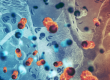
Dermatology is fundamentally a visual specialty; nearly every diagnosis we make is based on what we see. Yet historically, dermatology has often relied heavily on memory and narrative chart documentation rather than systematic photographic records of a patient’s skin. When a patient returns a year later and asks whether a mole has changed, physicians are frequently forced to rely on recollection, written descriptions, or the patient’s own perception.
One common approach in clinical practice is spot photography: taking photographs of individual lesions that appear atypical or warrant closer monitoring. Spot photography, often combined with dermoscopy, can be extremely helpful for tracking a specific lesion over time.
However, this approach has an inherent limitation. Spot photography only documents lesions that have already drawn attention. For patients with numerous nevi, it does not provide a comprehensive record of the entire skin surface. New lesions may appear in areas that were never photographed, and subtle changes may occur in lesions that were not previously identified as concerning. In effect, we may be carefully monitoring a small subset of lesions while the rest of the skin remains undocumented.
For patients with many moles, subtle change detection becomes a much more complex problem. During a routine skin exam, a dermatologist may visually assess dozens—or even hundreds—of lesions in a single visit. Expecting any physician, no matter how experienced, to perfectly remember the appearance and location of every lesion across multiple years is unrealistic.
Total body photography has long been recognized as a logical solution to this challenge. Capturing a complete, standardized photographic record of the skin at a baseline visit and then comparing it at follow-up should allow clinicians to identify new or changing lesions far more reliably. In a visual field like dermatology, systematic photographic documentation seems like an obvious step.
For most of dermatology’s history, however, the execution has been impractical. Skinmap represents a shift in that execution by turning a historically impractical concept into something that can function within the realities of a busy dermatology practice.
In my next blog post, I’ll walk through what has traditionally limited old-school total body photography and how Skinmap technology provides a practical, scalable solution in everyday clinical practice.